Pediatric dental management fibroma giant cell: case report
Keywords:
Fibroma, Células gigantes, DiastemaAbstract
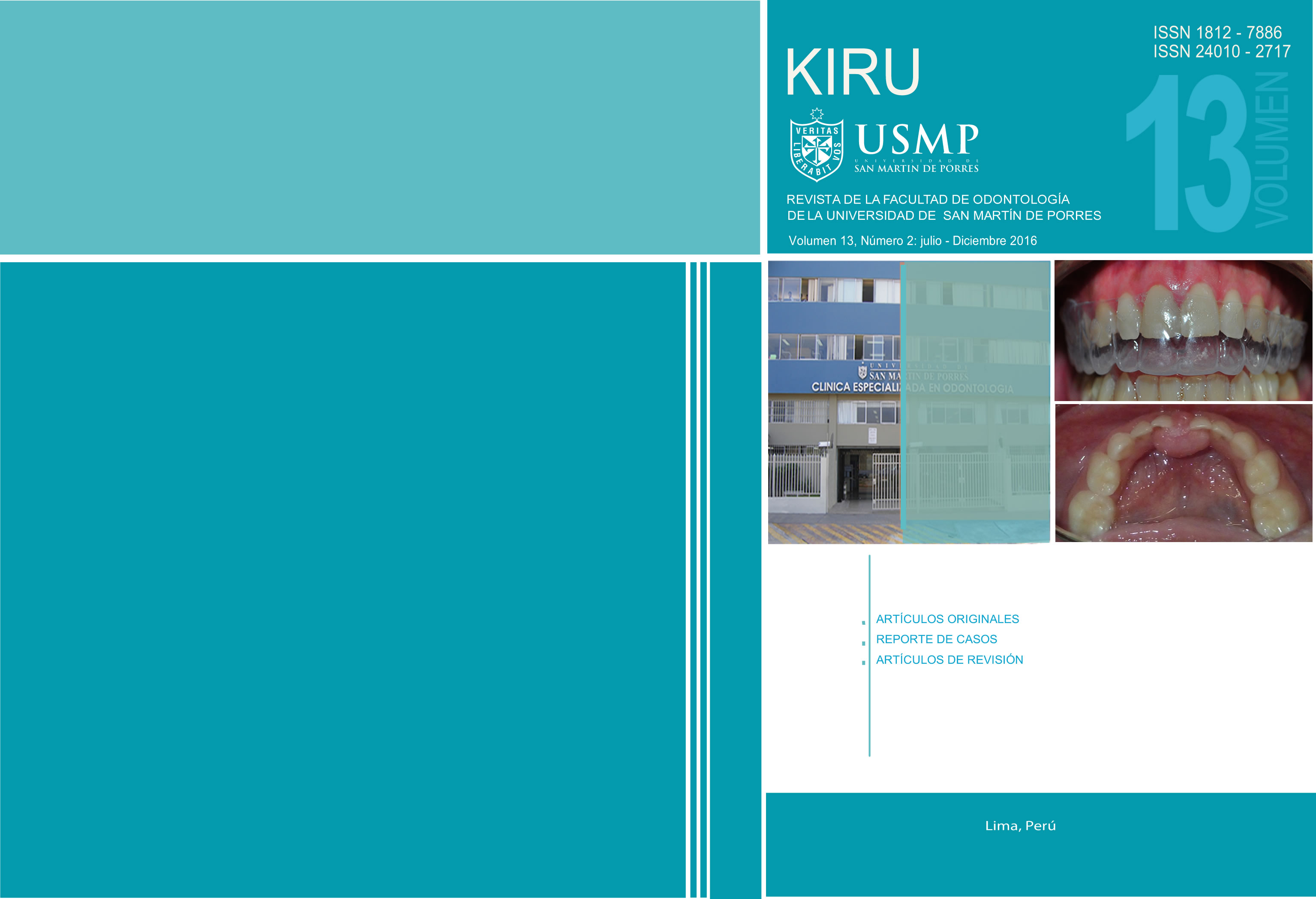
The present article, reports a clinical case of a female 37-month-old child, who arrived to the Specialized Dental Clinic of the Pediatric Dentistry Department from the University San Martin de Porres– Peru, at clinical examination the patient showed a soft tissue injury of about 1 cm in diameter, roundish, with smooth, shiny and well-defined limits and presenting a pedicle up to the deciduous lower incisors creating a diastema between the teeth. It was decided to realize surgical removal of the lesion to be a slow-growing lesion that is affecting speech and feeding the patient, determining the histopathology diagnosis of giant cell fibroma. The importance of correct diagnosis and treatment of these injuries, especially in the first years of life is something to highlighted.Downloads
References
Lukes S., Kuhnert J., Mangels M. Identification of a Giant Cell Fibroma. Journal of Dental Hygiene 2005; 79(3): 1 – 14.
Sekar B., Augustine D., Murali S. Giant Cell Fibroma - A Case Report With Review of Literature. Indian Journal of Dental Sciences. 2011; 3(5): 48 – 50.
Sampaio M., Domaneschi C., Daumas F., Weinfeld I. Giant Cell Fibroma of the Maxillary Gingiva in Children: A Case Report. J Dent Child 2010; 77: 174-6.
Vergotine R. A Giant Cell Fibroma and Focal Fibrous Hyperplasia in a Young Child: A Case Report. Case Rep Dent. 2012: 1-5.
Lukes S., Lautar Ch. Access. 2010; 24(10): 16-8.
Butchibabu K., Sushma N., Hussain W., Mishra A. Laser excision of a giant cell fibroma – report of a case and Review of literature. Aedj 2010; 2(4):221- 4.
Büchner A., Shnaiderman-Shapiro A., Mariiena Vered M. Pédiatric Localized Reactive Gingival Lesions: A Retrospective Study from Israel. Pediatr Dent 2010:32:486-92.
Neville, Brad W., Douglas D., Dean K. Color Atlas of Clinical Oral Pathology. 2003: 275-312.
Weathers D., Callihan M. Giant-cell fibroma. Oral Surg. Oral Med. Oral Pathol.1974; 37: 374.
Regezi J., Courtney R., Kerr D. Fibrous lesions of the skin and mucous membranes which contain stellate and multinucleated cells. Oral Surg. Oral Med, Oral Pathol. 1975; 39:197.
Houston. G. The giant-cell fibroma: A review of 464 cases. Oral Surg. Oral Med, Oral Pathol. 1982; 53: 582.
Odell E, Lock C, Lombardi T; Phenotypic characterisation of stellate and giant cells in giant cell fibroma by immunocytochemistry. J Oral Pathol Med 1994; 23: 284-7.
Eversole L., Rovin S. J oral Path. 1972; 1(1): 30- 8.
Büchner A., Shnaiderman-Shapiro A., Mariiena Vered M. Relative frequency of localized reactive hyperplastic lesions of the gingiva: a retrospective study of 1675 cases from Israel. J Oral Pathol Med. 2010; 39: 631–638.
Weathers D. Campbell G. “Ultrastructure of the giant cell fibroma of the oral mucosa,” Oral Surgery Oral Medicine and Oral Pathology. 1974; 38(4): 550–61.
Downloads
Published
Issue
Section
License
- Los autores/as conservarán sus derechos de autor y garantizarán a la revista el derecho de primera publicación de su obra, el cuál estará simultáneamente sujeto a la Licencia de reconocimiento de Creative Commons que permite a terceros compartir la obra siempre que se indique su autor y su primera publicación esta revista.
- Los autores/as podrán adoptar otros acuerdos de licencia no exclusiva de distribución de la versión de la obra publicada (p. ej.: depositarla en un archivo telemático institucional o publicarla en un volumen monográfico) siempre que se indique la publicación inicial en esta revista.
- Se permite y recomienda a los autores/as difundir su obra a través de Internet (p. ej.: en archivos telemáticos institucionales o en su página web) posterior al proceso de aprobación del manuscrito, lo cual puede producir intercambios interesantes y aumentar las citas de la obra publicada.


